We found 22515 price guide item(s) matching your search
There are 22515 lots that match your search criteria. Subscribe now to get instant access to the full price guide service.
Click here to subscribe- List
- Grid
-
22515 item(s)/page
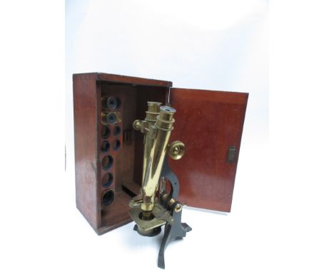


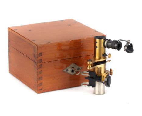
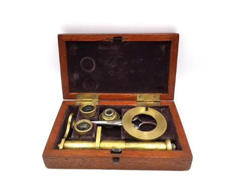
Lot 232
A Solar Microscope By Dollond, English, c.1790, engraved 'DOLLOND LONDON' the microscope of typical form with a brass plate with geared adjustment to rotation and angle of the mirror, with 2 large screws to fit to window shutter, main microscope body with 3 projection lenses in brass dovetails and a set of 6 magnifying lenses on a single dovetail, brass wet cell slider all in the original fitted mahogany case 27cm wide

Lot 1
Original Cast Aluminium Hospital Sign for 'PATHOLOGICAL LABORATORY', Original Cast Aluminium Hospital Sign for 'PATHOLOGICAL LABORATORY', with raised boarder and lettering, label to rear reads 'K. H. April 1994 from old block wall which was 100 years old', length 91cm Note what better sign to mount on a wall pointing to the room containing your microscope collection!!

Lot 229
Mid Victorian Binocular Microscope, Smith & Beck English, dated (from the Beck records) June 1862, engraved to the foot 'Smith & Beck, 6 Coleman Street, LONDON, 2408', the microscope on a equiaxial foot with tall upright column, plano-concave mirror in gimbal on sliding collar, focusing substage collar, mechanical stage with X-Y control, Lister limb with binocular body tubes, course focusing, screw fine focus with interocular adjustment at the top, in a fitted mahogany case with case of accessories including: 1 1/2" objective & can. 2/3" objective & can. 1/5" objective & can. 4/10" objective & can. pair of medium power eyepieces. Errector lens. Cased Maltwood finder. Compressor. Eyepiece micrometer. Achromatic condenser. Double nosepiece. Eyepiece lucida. Large livebox. Stage Forceps. 2 Leiberkuhns. Polariser. Analyser. Darkwells & Holder. Glass stages. and in the main case: Wenham Parabolic condenser. Pair of high power eyepieces. Monocular tube. Zoophyte Trough. Rotator & stops. Tabel condenser

Lot 233
A Zeiss IIa Compound Microscope, German, c.1890, engraved 'Carl Zeiss Jena No. 37534', microscope on 'Y' shaped base with plano-concave mirror, with Abbe substage illuminating apparatus, rotating stage, triple nosepiece, with 4 Zeiss objectives, 5 eyepieces all in fitted French polished mahogany case with leather outer case

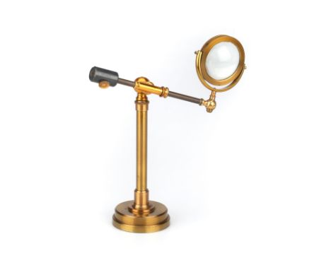
Lot 237
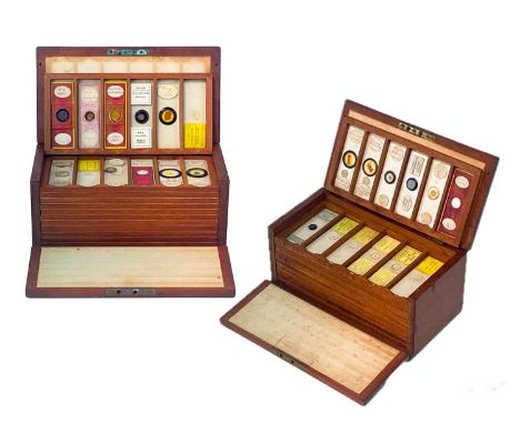
R & J Beck Microscope Lamp & Slide Preperation Outfit, A large 19th century French polished mahogany case with 8 drawers contining slide preperation equipment and a fitted compartment for a microscope lamp engraved aroun the base 'R & J BECK Ltd, LONDON', the lamp with circular brass base, square support, hand blown glass oil reservoir, with adjustable condenser lens and black chimney, case containing various bottels with labels for R & J Beck along with others, other preperation equipment, ringing tables, hand microtome and other equipment, case 33cm wide x 49cm tall

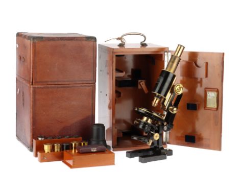
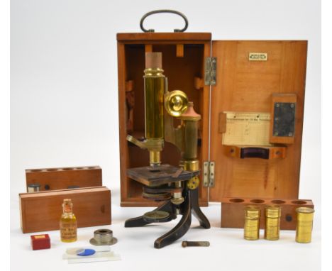
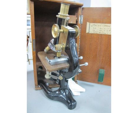
Lot 227
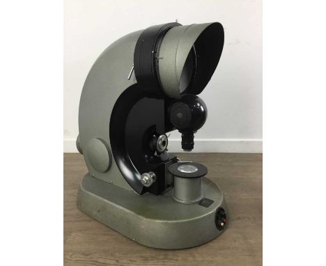
A Good Swift "Dick" Petrological Microscope English, c.1895, signed to foot ‘J Swift & Son, London’, standing on cast brass foot finished in black lacquer, trunnions at top support body, plano-concave mirror on gimbal below substage, substage assembly with rotating Nicol prism on rotating divided circle for angular measurement, square stage with Swift 2″ patent stage, main body to rear of stage incorporating the ‘Dick’ rotating mechanism with fine focus via screw and course focusing via diagonal rack work, body tube incorporating a sliding plate with aperture and slide in/out Bertrand lens, to top a rotating and folding analyser engraved with 45 degree positions, complete with 2 Swift objectives, 3 eyepieces in original mahogany case The Dick Petrographic Microscope by James Swift & Son: A Historical and Operational Overview Historical Context - The history of the "Dick" Petrographic Microscope is closely intertwined with the evolution of microscopy and mineralogical studies in the late 19th and early 20th centuries. This particular microscope was developed by James Swift & Son, a company that became a key player in the production of scientific instruments in England during that period. James Powell Swift initially worked under the instrument maker Andrew Ross before establishing his own company in 1854. As the company expanded and his son joined in 1877, it was renamed J. Swift & Son. By 1912, the firm had evolved into James Swift & Son Ltd. One of the significant advancements in the field of petrographic microscopy came from Allen B. Dick, an inventor who, in 1889, designed a unique gearing system that allowed for synchronized rotation of both the polarizer (beneath the stage) and the analyzer (above the stage). This innovation made it easier to observe and study mineral samples by eliminating the need for constant adjustment and re-centration of the specimen and objective lenses when rotating the stage. Swift and Son were the first to manufacture microscopes incorporating Dick's patented gearing system, and they introduced the first model in their 1891 catalog. Known as the "Dick Microscope," . Although it was expensive and relatively few examples remain today, it was used by prominent geologists and mineralogists, including during the British polar expeditions to Antarctica. In particular, photos from the expeditions show geologist Frank Debenham preparing samples using a Swift/Dick microscope, highlighting the instrument’s role in significant scientific research. Over the years, the Dick Microscope underwent several iterations, with various modifications made to improve its design and functionality. Although these microscopes were produced for many years, they were always considered premium instruments, and as a result, their numbers were limited. Today, surviving examples of the Dick Microscope are rare, and many are missing essential components such as the slotted eyepiece or waveplates. The Operation of the Dick Microscope The Dick Petrographic Microscope was designed for examining thin sections of minerals using polarized light, a key technique in petrographic analysis. The microscope’s construction allows for the observation of mineralogical structures in ways that are not possible with conventional optical microscopes. Key to its functionality is the polarizer, which sits beneath the stage, and the analyzer, positioned above the stage. These two components are crucial for creating "crossed polars," a method that significantly enhances the visibility of mineral structures by utilizing polarized light. Here’s how it works: 1. Polarized Light: When light passes through the polarizer, only waves vibrating in one direction are allowed through. When no sample is on the stage and the analyzer is aligned at 90 degrees to the polarizer, the field of view appears black—a condition known as "extinction." 2. Anisotropic Materials: When a mineral sample, specifically an anisotropic material (one that has different properties depending on direction), is placed on the stage, it alters the path of the polarized light. Instead of the black field seen during extinction, various colors or interference patterns appear, depending on the mineral’s optical properties and its orientation relative to the light. 3. Crossed Polars and Rotation: The Dick Microscope’s main innovation is the synchronized gearing mechanism that allows both the polarizer and analyzer to rotate together. This eliminates the need to manually rotate the stage and re-center the objective lens—a process that could be tedious and required great precision. With the polarizer and analyzer moving in unison, the specimen remains stationary, allowing for smooth and efficient analysis of even the smallest mineral grains. 4. Mineral Identification: By observing the way light interacts with the mineral as the polarizer and analyzer rotate, geologists can identify minerals based on their optical properties, such as birefringence, pleochroism, and extinction angles. This technique is especially useful for studying thin sections of rocks, where the optical properties of individual mineral grains provide clues to their composition and formation history. 5. Waveplates and Additional Features: Many petrographic microscopes, including the Dick model, were equipped with accessories like waveplates, which help to determine additional optical properties of minerals, such as their optical sign (positive or negative). However, many surviving examples of the Dick Microscope lack these additional features, possibly due to wear or loss over time. Significance in Geological Research - The Dick Microscope represents an importatn step in petrographic microscopy. By simplifying the process of rotating polarizers and analyzers, it facilitated the study of mineral structures, making it easier for geologists to carry out precise optical analysis. This design innovation became particularly valuable in the field of geology, where the accurate identification and analysis of minerals are essential for understanding rock formation and the Earth's history. The instrument's role in early 20th-century geological expeditions, such as those to Antarctica, underscores its importance in scientific discovery. The high precision and quality of the microscope made it a valuabel tool for researchers working in some of the most challenging environments on Earth. References - Bracegirdle, B. *Microscopes: A Short History*. - Powell, J. "The Evolution of the Petrographic Microscope." *Journal of Geological Sciences*, 1901.
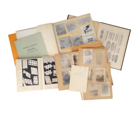
Lot 328
Book - Diatoms, Grove & Strut, Oamaru Deposits, & Others, hand written book by M.E. Parker, Lemington Spa 1956, concerning Oamaru diatoms from New Zealand, well anotated with many photomicrographs of diatoms and another book by the same titled 'DIATOMS a series of surplus prints from the collection made & photographed by J. W. Ryde Esq F.R.S. M.E. Parker. Leamington Spa July 1962 including a number of diatoms photographed in 1950 using General Electric Electron Microscope in 1950; a book on mounting diatoms by M.E. Parker, 1970 Other documents a copy of Diatomees, Especes Nouvelles Marines, Fossiles Ou Pelagiques par Jacq Brun, 1891;#
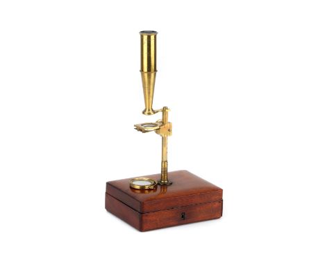
Lot 226
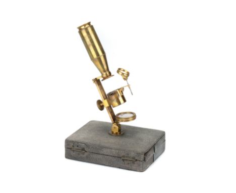
Georgian Compound & Simple Microscope Compendium, Englsh, c.1770, unsigned but similar in design to instruments built by John Cuff, the microscope when assembled fits inot a brass boss on the top of the lid, with rectangular sectioned support with hole at base for plano-concave mirror internally cut rack and pinion to focus stage, stage with internal sprung slide holder and lug to hold stage condenser, compound body tube, in fitted case covered in Ray skin lined with French silk velvet, complete with objectives and livebox, case 16.5cm
Lot 231
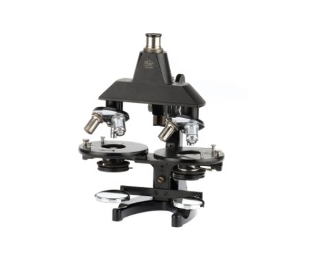
An Unusual Comparison Microscope By Zeiss, German, c.1930, engraved 'CARL ZEISS JENA Nr 305939' the microscope arranged on a 'U' shaped base with 2 identical microscope stages with Abbe type substage condensers, plano-concave mirrors, triple nose pieces with 2 sets of identical LOMO objectives, with beam splitting prism housing Note: possibly used by forensics for ballistics identification.
Lot 243
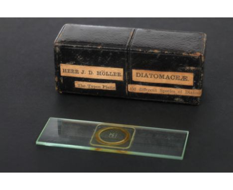
A Very Fine 400 Diatom Microscope Slide, Moller, German, dated 1870, slide engraved ‘Diatomaceen = Typen-Platte Deckglasdicke 1/9th mmtr, No.195, J. D. Moller Wedel in Holstein Germania 1870’ with four groups of diatoms in square arrangements, in a padded Moroccan leather case with labelling which reads ‘HERR J.D. MÖLLER DIATOMACEAE, The Typen Platte, 400 Different Species of Diatom’,all diatoms in place and in good condition
Lot 241
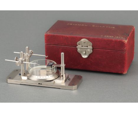
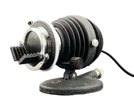
A Zeiss 'PRISMEN-ROTATOR' Microscope Accessory German, c.1890, signed in script 'Carl Zeiss Jena ', constructed of nickel plated brass with blued steel screws, engraved scale to rotator; in original leather covered wooden case, case width 12.8cm Note: This is the rare 'Double Prism Rotator' which incorporated a second prism to correct odd numbers of reflections, it appears in the Zeiss 1898 catalogue and cost an extra 18 marks more than the standard Prism Rotator, the description in the 1898 catalogue reads 'This apparatus is adapted for viewing from all sides, at low magnifications, large objects, in air or water, illuminated by incident light, without necessitating any alteration in the position of the object with respect to its support. This accessory will, accordingly, be found particularly useful for the examination from all sides of such objects as are too delicate or precious to admit of any preparation. By means of a rotating base and multiple reflections at silvered prism surfaces the object is optically placed in such a position that it may be examined from above or below, or any other point.'
Lot 234
A Large Zeiss Jug Handle Microscope, German, c.1900, engraved to the body tube 'CARL ZEISS JENA Nr. 43277' and engraved on the foot in script 'Katherine M. L. Gamge' then covered by another plaque (unscrewed) engraved 'Eric Impey, Cambridge' microscope with plano-concave mirror, Abbe condenser on rack focusing, rotating mechanical stage, single drawer tube, separate simple stage, 5 Zeiss objectives, 5 eyepieces in a Zeiss leather outer case
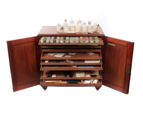
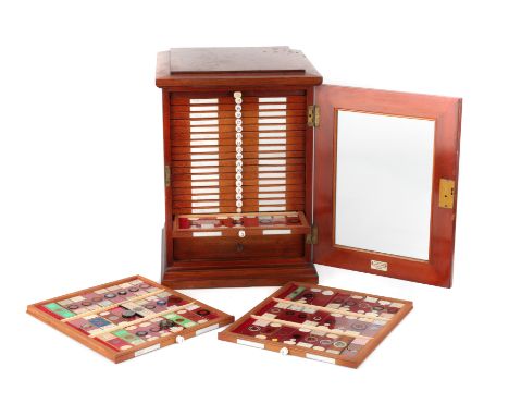
Lot 249
The Cabinet:English, c.1880, constructed of French polished mahogany, 2 doors opening to reveal a bank of drawers, label to inside of door reads 'ELCOCK - September 29th at 28 Fitzroy Avenue, Belfast, Harriet Sophia wife of Charles Elcock of a son, 1878', the top drawer containing a selection of slides, some by Elcock and others of diatoms and other subjects including one from the HMS Challenger Expedition, the bottom drawer containing a number of hand blown glass bottles and vials containing soundings from the HMS Challenger Expedition ( sounding dates, depths and locations corespond with the charts from 'VOYAGE OF THE CHALLENGER, WYVILLE THOMPSON, 1878', a copy of whihc is included in another lot), with 8 drawers of blank diatom slides and mounting equipment, the cabinet 59cm wide, 59cm tall, 33cm deep.Charles Elcock (1834-1910) was a well known figure in the world of microscopy, particularly known for his expertise in creating microscope slides featuring foraminifera, a group of amoeboid protists characterized by their intricate shell structures. Born in Pontefract, Yorkshire, England, on August 18, 1834, Elcock was the second son of Charles and Mary Ann Elcock. The Elcock family were Quakers, a religious background that profoundly influenced Charles throughout his life, as reflected in his writings and humanitarian efforts during the Franco-Prussian War. Elcock's early career was varied, encompassing teaching and publishing, but it was his later work in microscopy for whihc he is remembered.Early Life and Career.Elcock's early years were marked by a strong education and a diverse set of experiences. After the death of his father in 1837, his mother took up teaching to support the family. Elcock attended the Friends' School at Rawdon, which was a formative experience given the Quaker values emphasized there. His early professional life included roles as a teacher and printer, and he maintained a close association with the Quaker community throughout. By the 1860s, Elcock was involved in publishing religious texts, which eventually led to his work in London and Gloucester, where he likely developed his interest in microscopy through his connection with Alfred William Bennett, a prominent member of the Royal Microscopical Society.Microscopy and Foraminifera.Charles Elcock's most significant contribution to science was his work with microscopy, particularly in mounting foraminifera on microscope slides. Foraminifera are microscopic marine organisms that produce a shell, often referred to as a "test," which can be quite intricate and beautiful. Elcock's slides were celebrated not just for their scientific utility but also for their aesthetic appeal. His work involved arranging these tiny shells meticulously on slides, often organizing them by species and orientation, which made his slides valuable for both scientific study and as objects of beauty.Elcock's expertise in preparing these slides was widely recognized. He became a member of the Belfast Naturalists' Field Club shortly after moving to Ireland, where his skills in mounting foraminifera were lauded. In 1879, his work won a prize from the club for its artistic skill and superior finish. His techniques and methods were innovative, and he shared his knowledge through articles, most notably in the Journal of the Postal Microscopical Society, where he also advertised his slides.Legacy and Impact.Elcock's slides were distributed through well-known retailers in London, Manchester, and Bath, and they were highly regarded by contemporary scientists and hobbyists alike. Reviews of his work praised the meticulous attention to detail and the scientific value of the slides. His contributions to microscopy, particularly in the study of foraminifera, have left a lasting legacy in the field. While much of his life was also dedicated to religious writing and humanitarian efforts, it is his work in microscopy that has cemented his place in the history of science. A large part of his original equipment and the slides he produced is held at the Whipple Museum of Science in Cambridge: https://www.whipplemuseum.cam.ac.uk/explore-whipple-collections/microscopes/foraminifera-slides-and-working-tools-microscope-slide-maker Challenger Expedition: Revolutionizing Oceanography through Deep-Sea SoundingsThe Challenger Expedition (1872-1876), a pioneering oceanographic endeavor, marked a turning point in our understanding of marine sciences. This British voyage, named after the HMS Challenger, was the first dedicated scientific exploration to systematically study ocean basins, marine life, and geology. Among its most critical contributions were the extensive soundings, temperature recordings, and water samples taken during the expedition, which have had a lasting impact on oceanography.Deep-sea soundings, the process of measuring the depth of the ocean, were among the most revolutionary aspects of the Challenger Expedition. Utilizing newly developed sounding equipment, the expedition made nearly 500 soundings across the world’s oceans. These measurements were pivotal, not only in mapping the seabed but also in discovering the global patterns of oceanic trenches, underwater mountains, and plains.Prior to the Challenger Expedition, the depths of the oceans were largely unknown. The sounding techniques employed involved lowering weighted lines, known as sounding lines, into the ocean until they reached the seabed. The depths recorded by Challenger revealed for the first time the complex topography of the ocean floor. One of the most significant findings was the Challenger Deep in the Mariana Trench, recorded as the deepest part of the world's oceans.The data collected on these soundings provided foundational knowledge that spurred further scientific inquiry. For instance, the temperature profiles of ocean waters at different depths, also recorded during these soundings, helped scientists to begin understanding thermocline and its role in oceanic circulation patterns.The implications of these findings were vast. They challenged previous notions of a lifeless deep sea by providing evidence of life at all depths, and the samples of sediment helped develop the fields of marine geology and paleontology. This wealth of data collected by the Challenger laid the groundwork for modern oceanography and prompted the establishment of permanent oceanographic institutions.The Challenger Expedition was instrumental in transforming oceanography from a field cluttered with myths and speculations to a serious scientific discipline. Its soundings opened up new realms in the understanding of oceanic depths and laid down the benchmarks for future explorations, forever altering our relationship with the oceans. The expedition not only charted unknown waters but also set the course for future marine scientific endeavors, proving its legacy in the history of science.

Lot 246
Collection of Microscope Diatom Slides, Klaus Kemp Collection Collection of Group slides & Type Slides from the Collection of the Late Klaus Kemp. Strews by: K. R. Green, K. D. Kemp, Girodet, Rev. Vise, R. Pettigrew, A. Hepworth, R. Sutter, C. Baker, & F. Parrot. Type Slides by E. C. P. Bone, R. I Frith, L. N. Bramley J Tempere and others
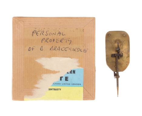
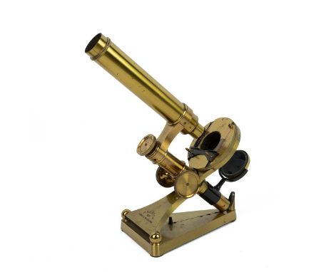
Lot 228
Brass Leeuwenhoek-type Simple Microscope, in the manner of the microscopes built by Maitland/Artis in the 19th century, the lens-plates 47.07mm x 27.6mm x 1.56mm with 3 rivets and hammer marks around lens aperture, with spherical lens, the long-screw adjuster with flattened handle, the shaped bridge-piece with vertical adjustment screw and focusing screw, with angle bracket and securing screw, good uniform patination, in a card case marked 'PERSONAL PROPERTY OF B. BRACEGIRDLE'Antonie van Leeuwenhoek (1632–1723), was a Dutch scientist and tradesman widely regarded as the "father of microbiology." His groundbreaking work with microscopes enabled him to explore a previously invisible world, revolutionizing the fields of biology and microbiology. Van Leeuwenhoek's life and scientific contributions were marked by curiosity, keen observational skills, and a commitment to scientific inquiry that led to some of the most significant discoveries in natural history. Born in Delft, Netherlands, Van Leeuwenhoek initially pursued a career in textile commerce. He worked as a draper, a profession that involved the inspection of cloth quality. His interest in lenses and magnification arose from his need to observe the threads of textiles more closely, a practice common in the industry. Van Leeuwenhoek’s advancements in microscopy were not the result of formal scientific training but rather the outcome of self-taught experimentation and dedication. While simple magnifying glasses had been in use for centuries, Van Leeuwenhoek’s microscopes were far more advanced. He did not invent the microscope, as early forms had been created by figures like Zacharias Janssen and Galileo Galilei. Instead, Van Leeuwenhoek’s unique contribution lay in his ability to improve the quality of lenses, enabling far greater magnification. Using small lenses, Van Leeuwenhoek built simple single-lens microscopes that could magnify objects up to 275 times, an unprecedented level at the time. His methods produced lenses of superior quality, allowing him to observe objects in finer detail than any other scientist of his era. Through his microscopic investigations, Van Leeuwenhoek became the first person to document and describe the existence of microorganisms, which he referred to as "animalcules." His observations included bacteria, protists, sperm cells, and red blood cells. In 1674, he reported his discovery of single-celled organisms living in water, and in subsequent years, he provided detailed descriptions of various forms of life, such as protozoa and algae. Van Leeuwenhoek’s observations of bacteria in dental plaque and other substances were particularly groundbreaking. His detailed descriptions of the minute organisms helped establish the foundation for microbiology. He meticulously recorded his findings in correspondence with the Royal Society of London, where his work was met with both skepticism and admiration. The impact of Van Leeuwenhoek’s work cannot be overstated. His discoveries transformed scientific understanding of life at a microscopic level and challenged prevailing notions of biology. Until his work, the existence of a microbial world was entirely unknown. His findings laid the groundwork for future scientists, such as Louis Pasteur and Robert Koch, whose work on germ theory and microbiology expanded upon Van Leeuwenhoek’s early observations. Although Van Leeuwenhoek did not publish formal scientific papers, he communicated his findings extensively through letters to the Royal Society, which translated and published them in *Philosophical Transactions*. His documentation and sketches of microorganisms were essential for validating his discoveries and spreading his influence across Europe. Additionally, his improvements to lens-making influenced the design of microscopes in subsequent centuries, establishing standards that would be refined in later technological advancements. Van Leeuwenhoek's contributions to science were widely recognized during his lifetime, and he continued his work well into old age. He was visited by notable figures such as Peter the Great of Russia, who was intrigued by his research. Despite his lack of formal education, Van Leeuwenhoek was elected a Fellow of the Royal Society in 1680. Brian Bracegirdle (1933–2019) was a well known figure in the fields of microscopy and the history of scientific instruments. His career began in the study of biology, where he earned a PhD in fungal spore dispersal from the University of London. However, his interests soon expanded into the history and development of microscopy, leading him to become a leading expert in the field. Bracegirdle had a long-standing association with the Science Museum in London, where he made significant contributions to its collections of historic microscopes. One of his key achievements was his work on cataloging and documenting historical microscopes, which helped preserve the legacy of this important scientific tool. His deep knowledge and passion for the subject were shared with a wider audience through his extensive writing. Bracegirdle was also a popular lecturer, and his engaging teaching style left a lasting impact on students and colleagues alike. Brian Bracegirdle authored numerous books on microscopy and related topics, many of which became essential reading for historians and scientists. His publications include A History of Microtechnique* (1978) Microscopes: A Short History* (1978) Beads of Glass: Leeuwenhoek and the Early Microscope (1983) Microscopical Mounts and Mounters* (1995), The Quekett Microscopical Club 1865–2015* (2016), and several volumes in the *An Atlas of Microscopy* series. These works have cemented his legacy as an authority in the history of scientific instruments. His contributions remain vital to our understanding of the development of microscopy and its role in advancing scientific knowledge.
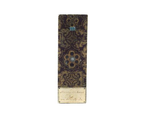
Lot 244
Farrant, R. J., 2 Micro Writing Microscope Slide, English, the slide signed at the bottom of the microwritting 'R. J. Farrants August 22nd 1855', with Farrant's recognisable paper to the front and rear with hand written label that reads 'Memorabel Events in 1/500 sq in', the engraveing reads: 1854 Jan 13 Combined English and French fleets enter the Black Sea March 12 Treaty of alliance between England, France and Turkey signed at Constantinople March 28 Declaration of War with Russia April 21 Bombardment of Odessa May 12 Loss of the Tiger steam frigate aground off Odessa Aug 16 Capture of Bomarsund by combined English and French fleets Sept 14 Allied English, French and Turks landed in the Crimea near Old Fort Sept 20 Battle of The Alma Oct 17 Attack on Sebastopol by the land batteries of the Allies and ships of the combined fleets Oct 25 Battle of Balaklava Nov 5 Battle of Inkerman Dec 13 Loss of the steamship Prince in storm in the Black Sea 1855 Feb 17 Russian attack on Eupatoria repulsed by the Turks May 24 Capture of ketch in the sea of Azoff by the Allies June 7 Assault and capture by the French of the Mamelou redoubt (Sebastopol) June 18 Combined French and English attack on the Malakhoff Tower and The Great Redan (Sebastopol) Aug 9 Bombardment of Siveaborg by combined English and French fleets Aug 16 Battle of the Tchernaya. Russians defeated by the French and Sardinians. R. J. Farrants August 22nd 1855
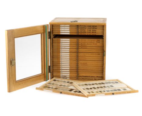
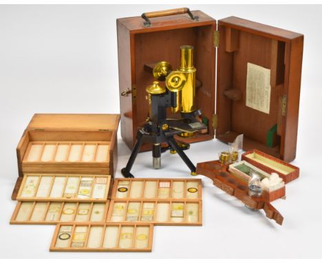
Lot 245
Large Victorian Microscope Slide Cabinet & Slides, the cabinet, English, c.1880, plaque to the inside of the door for 'W. WATSON & SONS MANUFACTURES 313 HIGH HOLBORN, LONDON', cabinet on plinth base with glazed door opening to reveal deep drawer for accessories (empty) with 22 drawers (one replacement) with ceramic labels, the cabinet contains a good mix of slides covering various subjects by many makers along with some amateur slides Footnote: This lot contains ivory and has been registered in accordance with the Ivory Act (Section 10), Ref.CS3GRBU2 Flints Auctions CANNOT ship this item out if the UK.
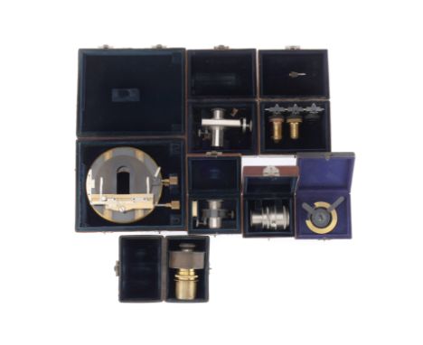
Lot 236
A Collection of Zeiss Microscope Accessories, including a large mechanical stage in a red leather case marked 'KREUTISCH 44 C. ZEISS JENA', a micrometer eyepiece in a leather case marked 'C. Zeiss Jena', a set of 2 of 3 objectives on quick change mounts in a leather case marked 'C. Zeiss Jena', a large eyepiece in a leather case marked 'C. Zeiss Jena', a large projection eyepiece with internal diaphragm in a leather case marked 'C. Zeiss Jena', a small eyepiece micrometer in a leather case marked 'C. Zeiss Jena', a condenser in a case marked 'C. Zeiss Jena',
Lot 224
Large Ross No.1 Compound Microscope, English, c.1854, engraved to the back of the foot 'A ROSS LONDON 567' the microscope standing on a massive 'Y' shaped base with tall uprights supporting the body of the microscope on large trunnions, with large plano-concave mirror on an articulated arm, substage with rack and pinion focusing, screw X-Y adjustment with early Ross (non RMS) diameter collar, stage with geared rotation, and X-Y control, large rectangular sectioned bar with bar limb on top with integeral fine focus mechanism compound body tube with objective engraved 'A Rofs 1854' and a single eyepiece
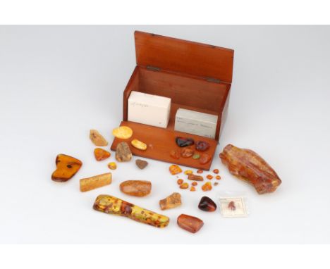
Lot 315
Collection of Amber Specimens, including 2 large specimens both containing a number of small insects, the largest 13cm long, a number of smaller pieces all containing inclusions and insects, along with 2 small card cases both containing small pieces of amber, all in an old microscope slide case
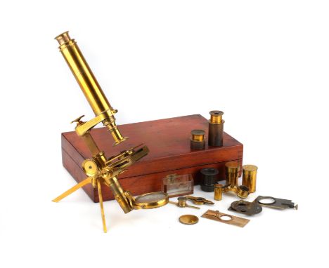
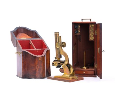
Lot 225
The First Folding Microscope by Smith & Beck, 1847/8, English, dated from the Beck records to late 1847/early 1848, engraved to the limb ‘Smith & Beck 6 Coleman St. London 177’ the microscope with folded tripod stand, large plano-concave mirror in gimbal on sliding collar, with folding mechanical Turrel-type stagecourse rack and pinion focusing, fine screw adjustment, in a flat fitted French polished mahogany case containing 2 objectives by Smith & Beck, 3 eyepieces, live boxes, zoophyte trough, polariser and analyser, Leiberkuhn, wheel of stops, Note: this model of folding travelling microscope is unrecorded in any of the Beck catalogues. No.177 is the first mention in the Beck sales books of a ‘Travelling’ microscope and was sold in early 1848 to J. D. Bagley. It was at this time that the firm Powell & Lealand released their design for a folding compound microscope around 1847. So it may have been made in competition or as a special one off that never made it into production. Either way this was, according to the Beck records the first folding microscope made by the company of Smith & Beck.

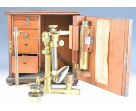
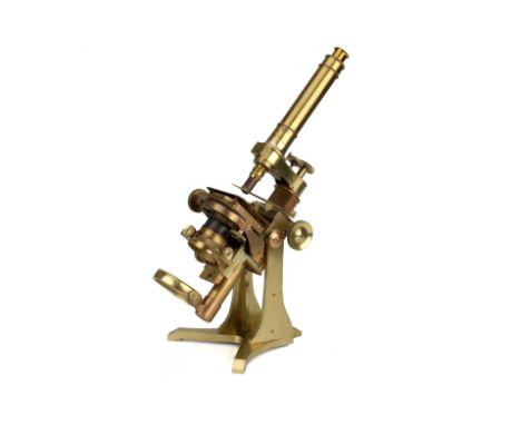
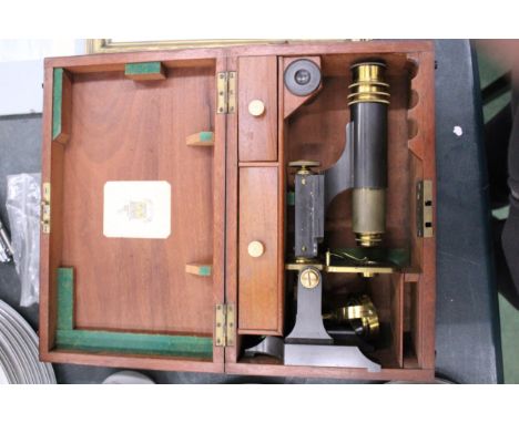
Lot 1242
Andrew Pritchard 263 Strand London 19th century brass microscope, in original flame mahogany carry case opening to reveal five graduated drawers with lenses and extensive selection of other accessories, some also named Pritchard and some Ross of London 33 Regent Street, height of microscope 47cm, with ivory licence
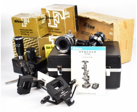
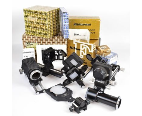
Lot 1435
Nikon accessories and attachments comprising PB-6E extension bellows, microscope adapter kit model 2, two F slide copy attachments, PB-6D spacer for PB-6 bellows, PB-6M macro copy stand, PS-4 slide copying adapter and two further sets of bellows, all but the last three items in original boxes

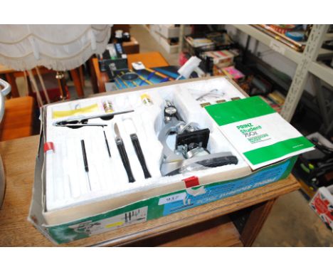

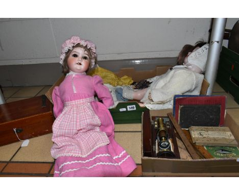
Lot 340
TWO BOXES AND LOOSE DOLLS, MICROSCOPE, WRITING SLOPE AND SUNDRY ITEMS, to include a Heubach Koppelsdorf 317.6 bisque head baby doll, with open mouth showing two teeth, brown glass sleeping eyes, composition body and limbs, an Armand Marseille bisque head doll, marked 'Armand Marseille, Germany, 390n, A 01/2 M' to the back of the head, with open mouth showing two teeth, blue glass sleeping eyes, composition body and limbs, a bisque head doll marked 'Special, Made in Germany', with open mouth showing two teeth (some teeth have broken off), brown glass sleeping eyes, composition body and limbs, with a selection of doll's clothing, together with a small brass microscope in a fitted case with instructions, eyepieces and five slides, a small writing slope with decorative metal strap details, approximate size 26.5cm x 17.5cm x 9.5cm, vintage maths books, rulers, etc (2 boxes + loose) (sd)


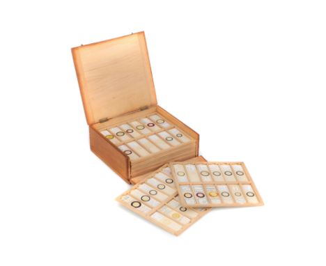
Lot 70
Two Victorian mahogany cases of microscope slides. The flush boxes, with hinged lids and fronts, containing 10 and 12 trays, the slides include several miniature photographs and images, including a £1000 Bank of England note, popular paintings, London, and other tourist attractions, many botanical and natural history subjects, geology, etc, approximately 120 slides, the boxes height 10cm, width 11.5cm, depth 21cm.
-
22515 item(s)/page